
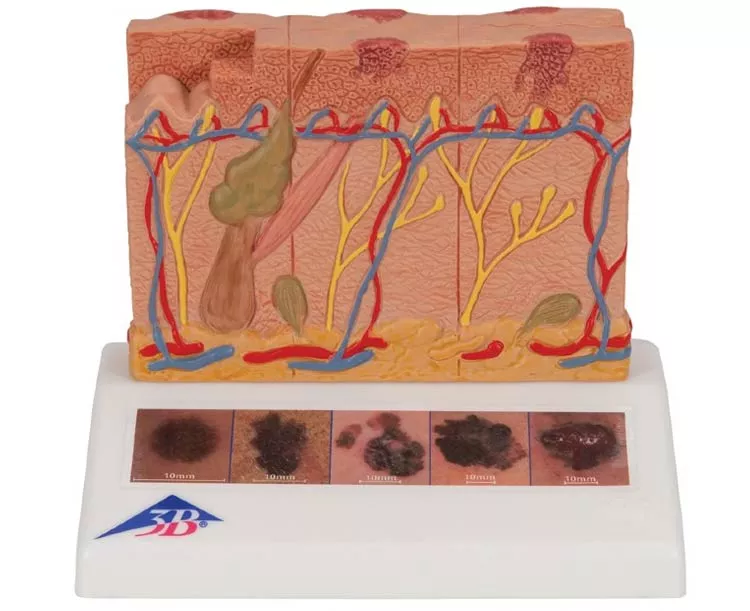
A great tool for illustrating this skin pathology, the Skin Cancer Stages Model by 3B Scientific shows healthy skin and 5 different stages of malignant melanoma on the front and back, enlarged 8 times:
- healthy
- malignant cells are found at the surface, within the epidermis
- malignant cells fill the epidermis, a few invade the papillary layer
- malignant cells fill the papillary layer
- malignant cells invade the reticular layer
- malignant cells have reached the subcutaneous fatty tissue, satellite cells approach a vein
In the top view of the skin cancer model, the individual stages of externally visible skin changes are shown, allowing for an assessment according to the ABCDE criteria. The sides of the skin cancer model show the various levels of invasion into the skin layers according to Clark (I-V) and the tumor thickness according to Breslow (in mm). 5 original color illustrations on the base of the skin cancer model show various types of malignant melanomas. The skin cancer model comes mounted on a base.
This anatomy model is manufactured in Germany.
Model No. J15